📗 -> 04/03/25: NPB173-L2
[Lecture Slide Link]
🎤 Vocab
❗ Unit and Larger Context
Small summary
✒️ -> Scratch Notes
The ultimate purpose of the brain and nervous system is to produce complex and adaptable movements
Neurons:
- conduct signals quickly over relatively long distances
- Action potential propogates
- Axons convey signals
- Synapses where axons terminate at junctions with soma or dendrites
- Neurotransmitters
Major cell types of nervous system:
- Glia
- Glia outnumber neurons. Their importance is not fully understood.
- Oligodendrocytes
- Astrocytes
- Microglia
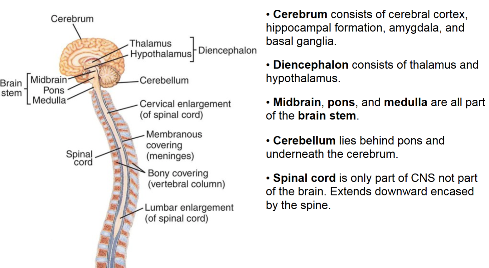
Nervous system has two parts:
- CNS and PNS
- Central and Peripheral nervous system
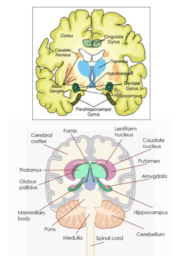
- CNS:
- Brain and spinal cord
- PNS:
- Nerves and ganglia outside the brain and spinal cord
- Importance of the distinction:
- Seperate developmental origin, BBB, different types of support cells, different response to injury
- At all division, the brain is bilaterally symmetric
Organization of the Spinal Cord
- Vertical segmental organization, with 31 in total
- Segments or divisions are associated with the entry/exit points of nerve roots between the vertebrae (bones of the spine)
- 5 major divisions, from top to bottom (or rostral to caudal apparently):
- cervical,
- thoracic
- lumbar
- sacral
- coccygeal
-
Each segment of the spinal cord is connected to the periphery with a left and right spinal nerve
- Nerves are bundles of axons
-
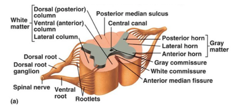
Each spinal nerve root has a dorsal (back side) and a ventral (belly side)
- Dorsal root: input, provides sensory info to CNS
- Ventral root: output, provides motor signals to the periphery
-
Cell bodies of primary sensory neurons are located in the dorsal rot ganglion
-
Gray matter (cell bodies) surrounded by white matter (nerve fibers)
- The gray matter is further separated into sensory and motor regions:
- Dorsal horn is mostly sensory
- Ventral horn is mostly motor
- Outer white matter of the spinal cord consists of distinct fiber tracts subserving different functions
- The gray matter is further separated into sensory and motor regions:
-
Total amount of white matter increases from the bottom (caudal), to the top (rostral) of the spinal cord
-
Amount of gray matter corresponds to musculature ()
Mapping between spinal cord segment and body location:
- The area of skin innervated by a single sensory nerve is called a dermatome
- The face is not innervated via the spinal cord, instead innervated by the cranial nerves
- Mostly ordered from rostral to caudal along the spinal cord (makes more sense thinking about distribution as a four legged animal rather than human)
- The group of muscles innervated by a single nerve is called a myotome
This structure allows deduction of nature of damage
Spinal Cord Injuries:
- 6 million people worldwide, around 40 new cases in the US per year per million people
- Most common causes are vehicular accidents, falls, violence (gunshots), and sports injuries. Mostly external trauma
- Can also be due internal factors, tumors or ischemia (inadequate blood supply)
- Can lead to partial or complete loss of motor and/or sensory function in the affected body regions (paralysis)
Timecourse:
- Spinal cord injuries most ofen result from compression, stretching, or laceration (cutting)
- Most traumatic injuries have two components, a primary and secondary
- Primary: Refers to the direct result occurring immedialy at time of initial trauma. This can include severing of axons, death of neurons and glia, and damage to the vasculature
- Circumscribed damage to neural tissue or other abnormalities are called lesions
- Secondary: Indirect after effects from processes initiated by the trauma.
- Can be as serious as the primary components: traumatic brain injury, a substantial proportion of deaths occur to secondary components unlike other forms of trauma
- Three phases of secondary spinal cord injury: acute, intermediate, and chronic
- Acute: The glial response to the primary injury initiates scar formation. Cut and crushed axons also begin to degenerate distal (away from the cell body) to the site of unjury (Wallerian degeneration)
- Intermediate and Chronic Phases: Axonal regeneration is limited
Wallerian Degeneration:
- Primary: Refers to the direct result occurring immedialy at time of initial trauma. This can include severing of axons, death of neurons and glia, and damage to the vasculature
- An active response to axonal injury.
- Involves changes in both axons and glial cells along the axons.
- Begins 1-2 days after injury
- Axonal skeleton disintegrates and axonal membrane breaks apart distal to the injury
- Followed by loss of the myelin sheath that surrounded the former axons
- Finally, macrophages clean up the mess
Myelination:
- Provided by different cells in CNS and PNS:
- CNS: Oligondendrocytes, provide multiple segments of myelin and can ensheath many axons
- PNS: Schwann cell provides only a single segment of myelin, only partially sheaths one axon.
- Supports axonal regeneration!
Regeneration in PNS
- Supports axonal regeneration!
- At the end of Wallerian Degen, the axon forms its growth cone, a specialized structure that promotes axon extension
- Route finding usually directed by extracellular environment, but factors used during development are lacking in adulthood
- Schwann cells re-align to form hollow tubes along the previous route of the axon
- Axon growth proceeds through these tubes until the original target is re-innervated
Oligondendrocytes can’t do this!
Spinal Cord Pathways:
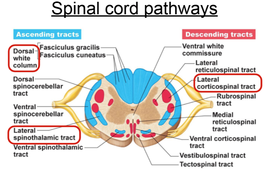
- Tracts in the white matter
- Tracts organized by location, not haphazard
- Descending:
- Subserve motor pathways from the brain to the periphery
- Ascending:
- Subserve sensory pathways from the periphery to the brain
Ascending:
- Dorsal white column
- Lateral spinothalamic tract
Descending: - Lateral corticospinal
Major Pathways
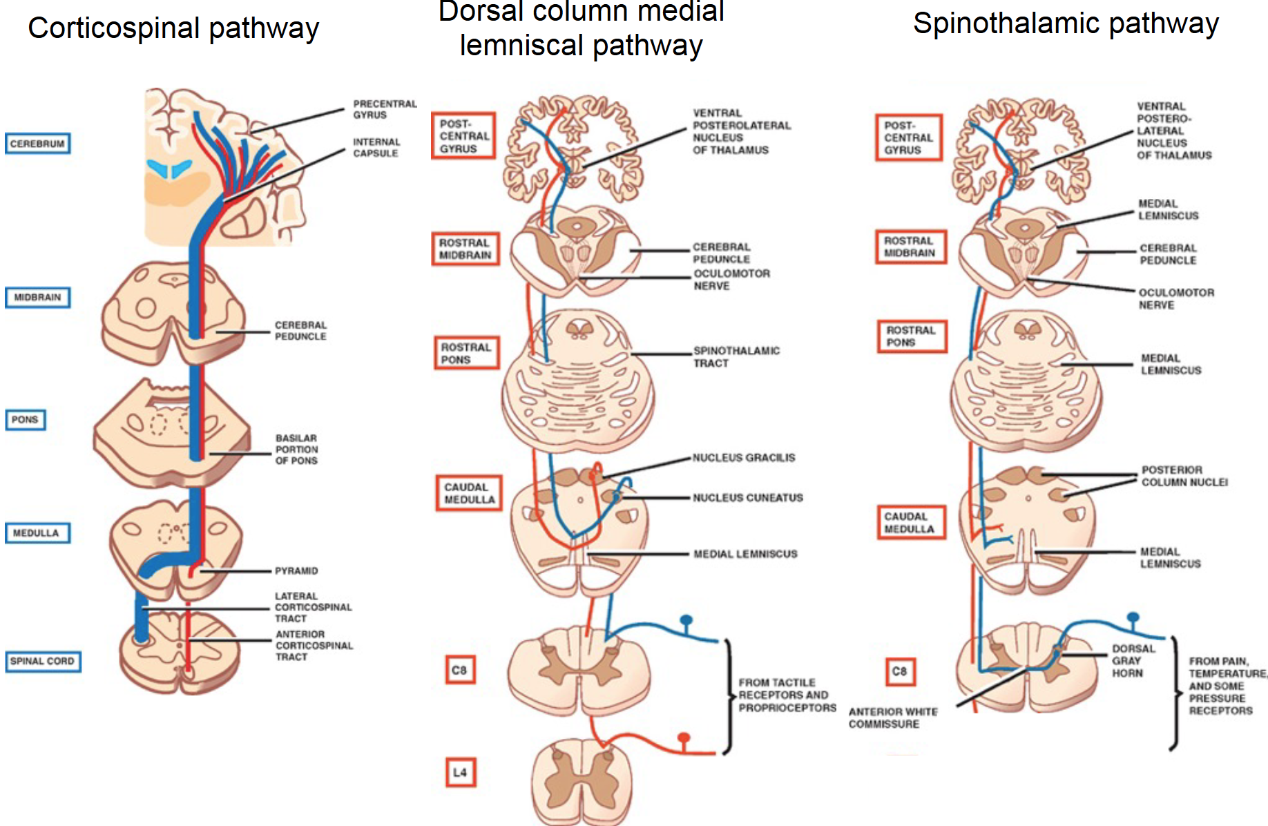
Spinothalamic (anterolateral) pathway
- Carries somatosensory signals
- First order neurons are connected to pain and temperature receptors
- First order neurons have cell bodies in the dorsal root ganglia
- Second order neurons send axons that immediately cross the midline (decussate)
- Second order neurons travel up the spinal cord in the anterolateral part of the white matter
- Second order neurons project to neurons in the thalamus
- Third order neurons project from the thalamus to somatosensory cortex
Dorsal column medial lemniscal pathway - DCML
- Also carries somatosensory signals
- First-order neurons are connected to touch and proprioceptive receptors
- First order neurons have cell bodies in the dorsal root ganglia
- First order neurons project up the dorsal columns to neurons in the medulla on the same side of body as their receptors
- Second order neurons send axons that immediately cross the midline (decussate)
- Second order neurons travel up the medial lemniscus
- Second neurons project to neurons in the thalamus
- Third order neurons project from the thalamus to somatosensory cortex
Lateral Corticospinal (pyramidal) pathway
- Lateral corticospinal pathway carries motor signals (mostly for limbs)
- Upper motor neurons have cell bodies in cortex (M1)
- Upper motor neuron axons immediately cross the midline before entering the spinal cord and don’t cross again
- Thus axons are on the same side of the muscles they control throughout the length they travel in the spinal cord
- Upper motor neuron axons project in the lateral column of the spinal cord to lower motor neurons in the ventral horn
- Lower motor neuron axons project throughout the ventral root to directly innervate muscles on the same side
Learning objectives
- Basic function of neurons and glia.
- Large-scale division of the nervous system.
- Components of white matter vs. gray matter.
- Segmented structure of spinal cord.
- How dermatomes reveal vertical organization of spinal cord.
- Dorsal vs. ventral organization of the spinal cord.
- Wallerian degeneration.
- Axonal regeneration and differences between PNS and CNS.
- Deductive approach for inferring spinal injury based on symptoms and vice-versa.
Questions:
What is cranial shift?
What musculature is he talking about in the spinal cord gray matter slide?
Dermatome vs myotome?
🧪 -> Refresh the Info
Did you generally find the overall content understandable or compelling or relevant or not, and why, or which aspects of the reading were most novel or challenging for you and which aspects were most familiar or straightforward?)
Did a specific aspect of the reading raise questions for you or relate to other ideas and findings you’ve encountered, or are there other related issues you wish had been covered?)
🔗 -> Links
Resources
- Put useful links here