Week 1 - Intro & Techniques
// Review Slides:
Measuring Neural Activity
- Invasive
- Extracellular Recording
- Single-unit
- LFPs
- Multiple contacts electrode
- Multiple electrode array
- Intracellular recording
- iEEG
- Optical Imaging (intrinsic signal optical imaging, VSD, CaSD)
- Non-invasive
- EEG, MEG, fMRI
Manipulating neuronal activity- Inactivation
- Lesions
- Patients with lesions
- Experimental lesions
- Reversible inactivation
- Muscimol injection
- Cooling
- Optogenetic stimulation
- Activation
- Microstimulation
- Optogenetic stimulation
Intro:
- Systems Neuroscience at different Levels
- Systems Approach
Techniques:
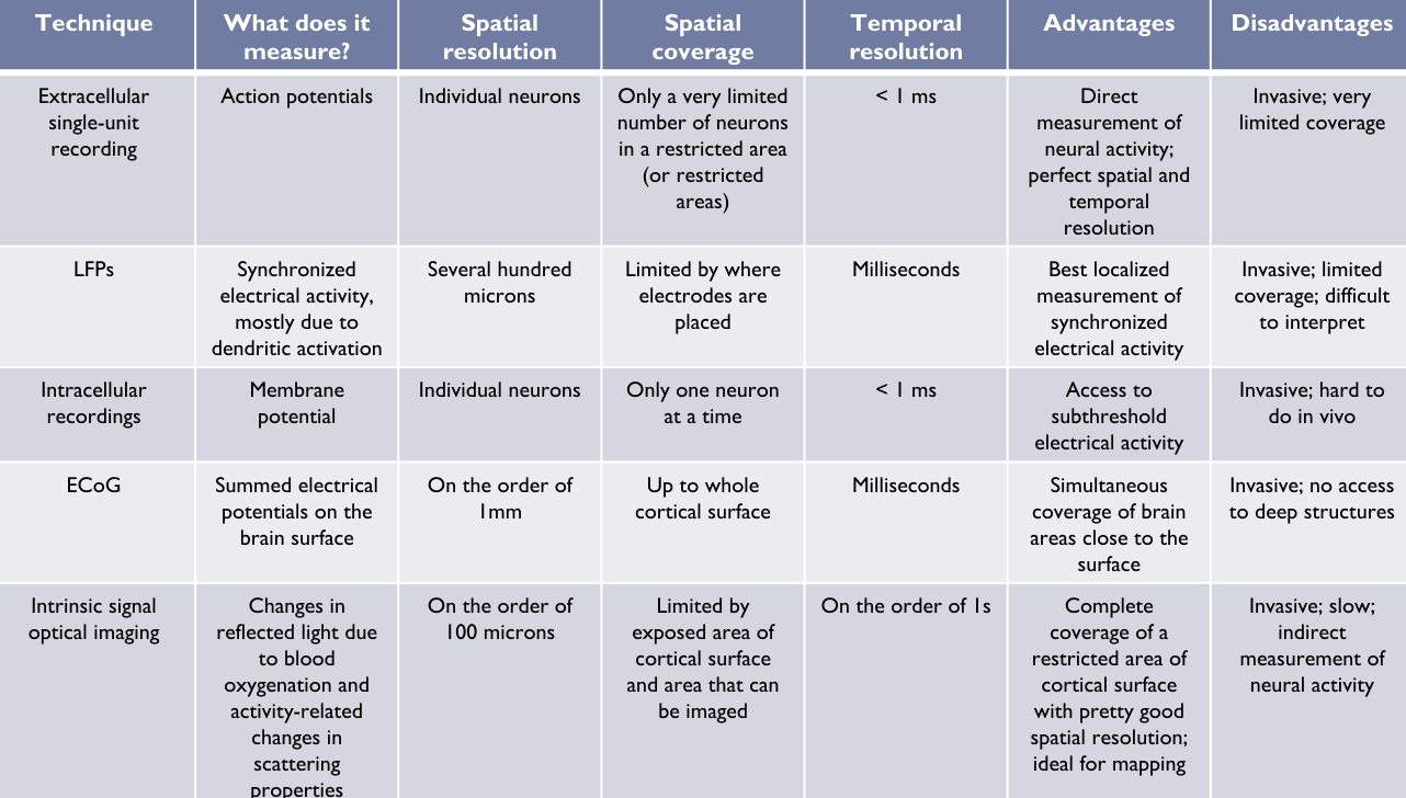
Invasive
Extracellular Recordings - Electrodes with a metal core recording from neural tissue. Only the sharp tip exposed, voltage between electrode and reference measured.
Single Unit Recording - Electrode tip placed close enough to measure from a individual neuron. Strength of recording depends on:
- Distance from soma
- Size of neuron
- Subject to sampling bias (excitatory bigger for example, and recordings more biased to sample bigger spikes)
- Can be used to build Raster plots and PSTHs
Local Field Potentials - Aggregations of neural activity (on scale of several hundred microns). Difficult to interpret.
Multi Contact Electrodes - Tetrodes / Multi contact rod - Multiple contacts help to seperate signals from multiple neurons
- Neuropixels probe (1,000 contacts)
Multi Electrode Arrays - Utah array
Intracellular - Records INSIDE membrane. Can record subthreshold EPSPs and IPSPs. - Very difficult to do
iEEG / ECoG - EEG directly on cortical surface - Simulatenous recording of LFP-like signals
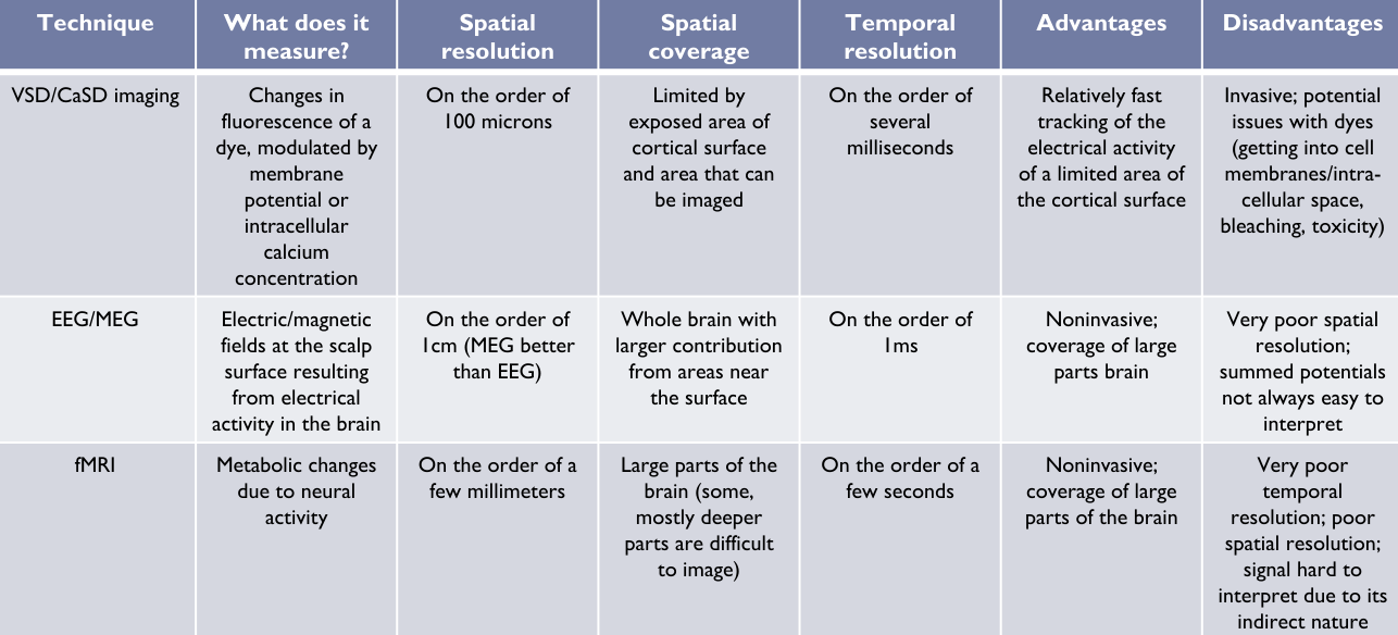
Optical Imaging - Use a camera to see how brain changes with certain dyes. - Voltage sensitive (VSD) / Calcium sensitive (CaSD)
Non-invasive
EEG - duh
MEG -
fMRI -
Manipulation
Quick Summary
- Inactivation
- Lesions patients with lesions experimental lesions
- Reversible inactivation Muscimol injection Cooling
- Optogenetic stimulation
- Activation
- Microstimulation
- Optogenetic stimulation
Techniques
Lesions
Reversible Inactivation
Microstimulation - Stimulate a small area with a extracellular electrode.
- around scale of a few hundred microns
- Acts fast (can cause stim within 5ms)
- to have a predictable effects needs to be applied in an area of similar neurons (so that effects are predictable)
Optogenetic - Introduce light dependent molecules into cell membrane to depolarize or hyperpolarize the neuron - Specific cells can be targeted
Week 2 - Neural Codes
// Review Slides:
- Single neuron rate code
- Population rate code (place code vs value code)
- Noise correlation, signal correlation
- Information theory (entropy, mutual information)
- Single neuron timing code
- Temporal population code
- Generating population response (spike generator, Poisson process)
- Reading out population response (population vector decoder, maximum likelihood decorder)
- Entropy and mutual information
Week 3-6 - Sensory Systems
// Review Slides:
Visual System
- Visual sensory epithelium: Retina (Rod, Cone, center-surround RFs)
- LGN (retinotopic maps)
- V1 (simple, complex cells, depth perception-disparity, RF mapping)
- Retinotopic map, oriental preference map, ocular dominance map
- Visual processing beyond V1
- Ventral pathway (V2: illusory contour, border ownership; V4: color consistency; IT: object recognition)
- Dorsal pathway (motion processing, MT, MST)
Auditory System
- Auditory sensory epithelium: organ of Corti
- Cochlea (cochlear hair cells, frequency tuning)
- Sound localization: superior olivary nuclei, ITD, ILD
- A1
- Tonotopic map
- Spatial tuning (horizontal location)
- Synchronized code: low frequency & rate code: high frequency
- Cortical analysis of complex sound
Sensory Systems - Feedback, predictive coding, anticipating sensory consequences of own actions
- Predictive coding
- Anticipating sensory consequences of own actions
- Re-afference vs “efference copy” / “corollary discharge”
- Neural activity in sensory areas affected by predicted sensory consequences of actions (e.g. V1, MST)
Week 6-8 - Motor Systems
// Review Includes
Oculomotor System
Skeletal Motor System
// Review Slides:
Oculomotor System
- Purpose of eye movements
- Six types of eye movements
- Saccade, pursuit, VOR, optokietic movements, vergence, fixation
- Eye rotation and extraocular muscles
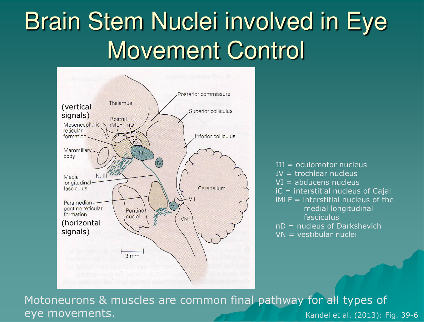
- Brain stem nuclei (oculomotor integrator circuitry)
- Superior colliculus
- Saccade generation (SC, FEF)
- Smooth pursuit
- VOR
Motor 1: Motor Systems Intro
Motor 2: Oculomotor System
6 Types of Eye Movements:
Conjugate vs. Disconjugate:
- Conjugate: Relies on eyes agreeing, working together to compile info
- Disconjugate: Seperate, relying on disparities in retinal position etc.
- Saccade (conjugate): fast motion to get objects of interest into fovea
- No visual feedback during motion
- reflexive, automatic response to visuals
- Smooth Pursuit (conjugate) - Stabilize a moving object in fovea
- Driven by moving stimulus
- VOR (vestibulo-ocular reflex; conjugate) - Stabilize still image while head is moving quickly
- Driven by vestibular input
- Optokinetic movements (conjugate) - Stabilize still image while head is moving slowly
- Driven by vestibular input
- Vergence (disconjugate) - Align images of near/far objects in both images
- 3 systems?? accomodation, vergence, pupillary constriction
- Not explained in slides, but:
- Motor systems to control how light hits your eyes retinas
- driven by retinal disparity
- 3 systems?? accomodation, vergence, pupillary constriction
- Fixation - Keep eyes on an object
- Active suppression of unwanted eye movements
Brain Stem Nuclei
Saccade Generation
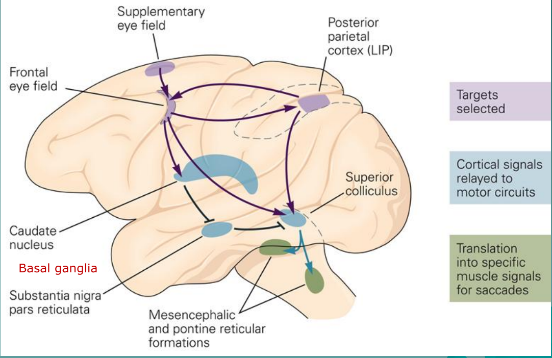
Major Players
- Targets Selected: PPC (LIP), SEF, FEF
- Cortical -> Motor: SC, SNPR, CN
- Motor -> Muscle: MG/PPRF
SC and FEF are the only areas that can send signals to the muscles. With them BOTH lesioned, saccades will disappear.
Horizontal Saccades
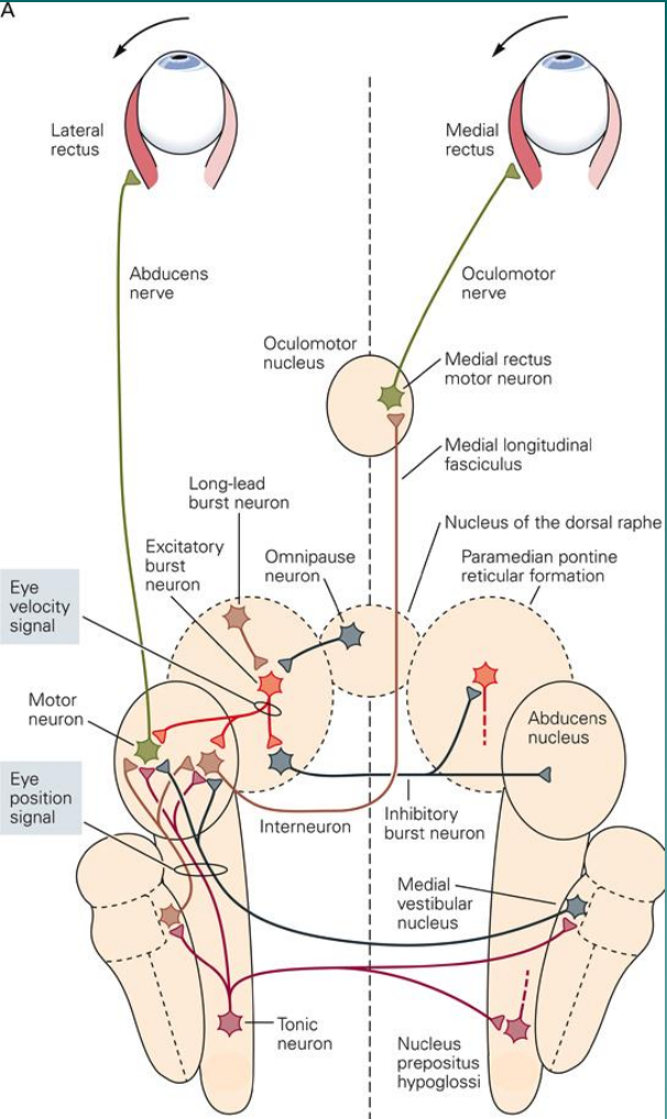
Summary of Players:
-
Eye: Final target, makes the saccades
-
OM (oculomotor nucleus)
- Excites inner eye muscle (medial rectus)
- medial rectus moro neuron
-
NOR (nucleus of the dorsal raphe):
- Omnipause neuron
- Holds eye position stead
-
PPRF (paramedian pontine reticular formation):
- long-lead + excitatory burst neurons
- Inhibitory signal
- Inhibitory signal to contralateral AN motor neurons
- Excitatory to ipsilateral AN
-
AN (abducens nucleus)
- motor neurons activate ipsi eye’s lateral rectus
- interneuron which excites medial rectus motor neuron
-
MVN (medial vestibular nucleus)
-
NPH (nucleus prepositus hypoglossi)
- Tonic neuron: keeps eye in same position
-
The eye receives input from:
- Abducens Nucleus (AN) (excites outside)
- Oculomotor (OM) nucleus (excites inside)
-
OM only receives input from AN
-
AN receives input from:
- Paramedian Pontine Reticular Formation (PPRF)
- Medial Vestibular nucleus (MVN)
-
NPH are inhibitory
Transclude of NPB163-Final-Prep-2025-06-07-18.32.20.excalidraw
Integrator Problem
Need to maintain current level of activity in order to maintain activity
-
- recurrent excitation helps to MAINTAIN activity (keep at 50%)
-
- mutual inhibition helps to COORDINATE activity (keep both sides in stride)
Smooth Pursuit
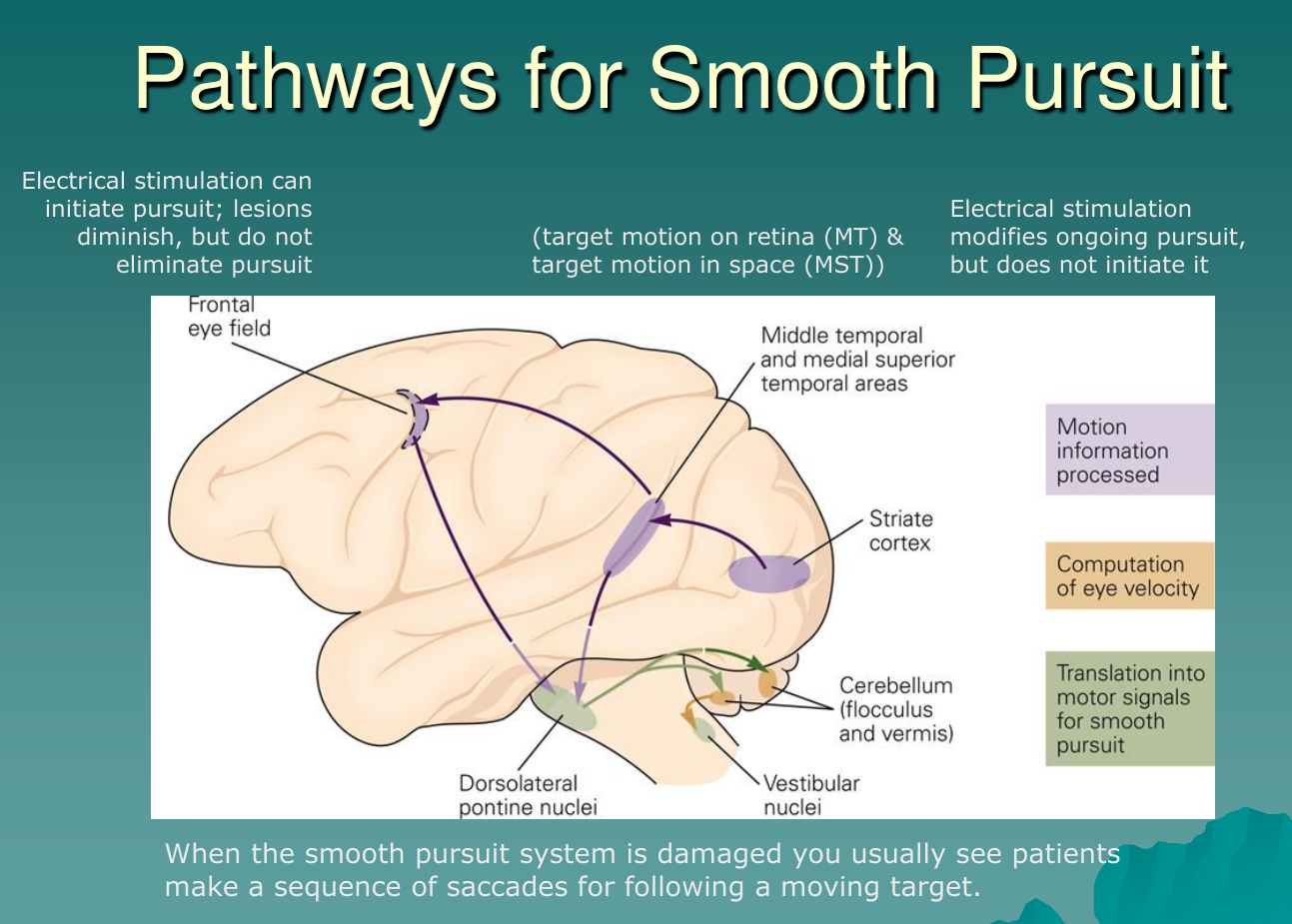
- Process Motion: FEF, MT/MST, GP
- Comp Vel: Cerebellum (flocculus and vernis)
- Signal -> Motor: DPN, Vestibular nuc
VOR - Vestibulo Ocular Reflex

Transclude of NPB163-Final-Prep-2025-06-07-19.14.45.excalidraw
Major Players
VOR:
- Built for very small delay: endolymph -> VN -> motor pathway
- fast rational acceleration only
small tracking handled by the optokinetic pathway
- “responds to very slow motion, but builds up slowly”
VOR & optokinetic both stabilize an image while the head is moving
- VOR: fast motion -> small/little delay
- Optokinetic: slow motion -> large time to build up
Motor 3 & 4: Skeletal Motor System
Skeletal Motor System
- Skeletal motor system
- Motor unit
- Spinal reflexes
- Properties
- Types of reflex (e.g. withdrawal reflex; stretch reflex)
- Reflex vs. voluntary movements
- Descending pathways
- Cortical movement control
- Cortical movement control areas (cortical spinal connections)
- M1 organization
- Functions of M1:
- Encode movement parameters
- Optimal feedback control theory
- Graziano experiment: map of behaviorally relevant postures
- Functions of PM, SMA
- Optimal motor control theory
Motor 3
Overall Pathway
Includes M1, Basal Ganglia, Cerebellum, and spinal cord
Motor neurons in the spinal cord are final common pathway for all motor action
Motor Unit - Muscle fiber &
One to Many: One motoneuron innervates Many (35-2,000) muscle fibers
- One muscle fiber can only be innervated by one muscle fiber
Columnar Organization
a-Motoneurons innervating a particular muscle form a column in the spinal cord, can span several segments
Proximal-distal rule:
- Medial motor neurons project to more proximal muscles (close to body?)
- Lateral motor neurons project to more distal muscles (farther from body)
- Somatotopy at all levels
Motor Units
Receive input from:
- Descending pathways (- and +)
- Peripheral input (+)
- Interneuron (-)
Outputs: - To muscle fibers at neuromuscular junction (nmj)
- Releases acetylcholine (ACh) into synaptic cleft
Recruitment
- Happens in a fixed order from weakest to strongest (use smallest force necessary)
- Deactiation follows reversed fibers
- Stronger muscles fatigue more quickly
- Smaller motor neurons (MN) innervate weaker fibers, larger MN -> stronger fibers
- Same excitatory current from spinal interneuron causes a larger EPSP in the smaller motor neuron
Spinal Reflexes
Simplest sensorimotor circuits
Contained all within SC
- Provided by receptors in muscles, joints, and skin
Reflexes are also more complex than usually given credit: - can adapt to the task
- not all or nothing, spatial extentnt and force are graded
Protective functional (withdrawal) - In upper extremities: withdrawal affects only one limb
- In lower: affects both limbs due to their role in stabilization (balance)
- General principle: reciprocal innvervation (activation of agonist and deactivation of antagonist)
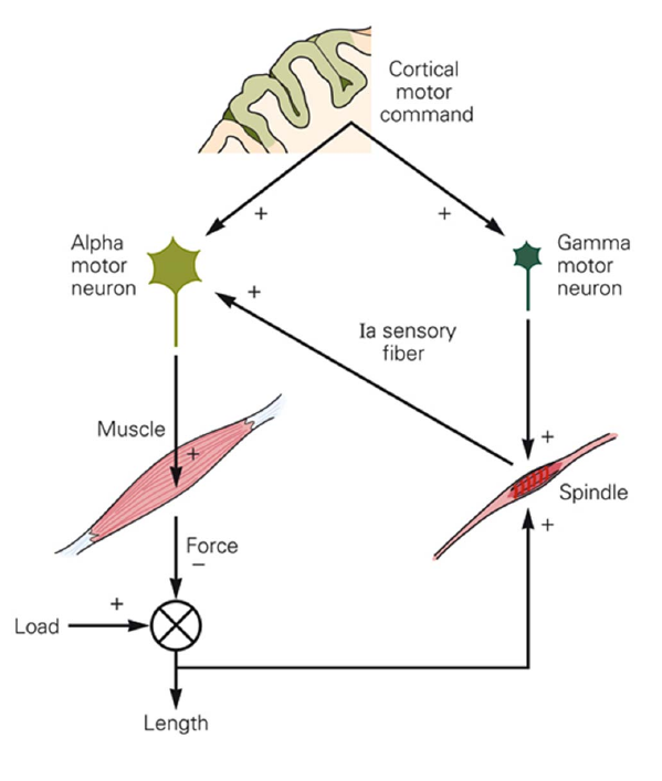
Muscle Spindles
Length sensors - Gives feedback corresponding to muscle stretch. Greater stretch/contraction -> Greater activation
Gamma motor neurons - Tune the sensitivity of muscle spindles. An example of predictive coding
- Adjusting the operating range of the muscle spindles
- In many movements alpha and gamma motor neurons activate in parallel, and “expect” the stretch from voluntary movements
Stretch Reflex
Back to reciprocal innervation, afferent stimulation from the:
- extensor nerve is excitatory towards extensor motor neurons
- flexor nerve is inhibitory towards extensor motor neurons (there is an inhibitory interneuron, flipping signal)
However, its more complicated than this and reciprocal inhbition can be blocked if co-contraction is desired (stiffening joints)
Golgi tendon organ
Signal tension in a muscle
- Protective function, but the organ also provides precise information about the state of contraction of a muscle
Projects to 1b inhibitory interneurons, who receive their principal input from the Golgi tendon organs
Reflexes and Voluntary Movements
The reflex circuits are not silent during voluntary movement.
They are part of a larger control loop, and they both leverage motor machinery
- Alpha and Gamma motor neurons both leveraged from cortical systems
Descending Pathways
Again the medial/lateral rule applies:
- Medial descending systems contribute to control of posture
- Lateral descending systems
- cat/monkeys: rubrospinal tract important for goal directed movements especially of arm and hand
- apes/humans: corticospinal tract (cst) assumes control of these functions
Brian stem integrates spinal reflexes into a variety of automated movements that control posture and locomotion
Descending motor pathways (in humans):
- Mainly terminate on spinal interneurons
- Only certain corticospinal fibers (esp for arm and hand movements) synapse directly onto motoneurons
- All descending pathways (EXCEPT the corticospinal tract) remain ipsilateral
- 90% of corticospinal fibers cross
- Vestibulospinal and modullary/pontine reticulospinal tracts: posture control
- Vestibulo -> Balance, so posture
- medullary / pontinue reculospinal -> ?
- Tectroreticulospinal tract: orienting head movements (signals originate in the SC)
- Corticospinal tract (cst): control of precise movements of distal extremities (signals originate in pyramidal cells in motor cortex)
Pneuomic:
Lateral Tracts:
- Corticospinal tract (CST), specifically lateral portion
- Rubrospinal tract
Medial tracts:- Everything else…
- Vestibulospinal and modullary/pontine reticulospinal tracts
- Tectroreticulospinal tract:
- Ventral corticospinal tract (CST)
Motor 4
Motor Areas (again)
Motor areas in FC, basal ganglia, cerebellum, brain stem, spinal cord
Cortical Connection
- Inputs to primary motor cortex:
- SMA, PMd, PMv, S1, BA5?
- Inputs to premotor areas:
- PFC, BA5, BA7
Corticospinal Connections
- PFC, BA5, BA7
- Betz cells (large pyramidal cells in layer 5 of M1) thought to be important for cortico spinal fibers
- Actually we think that most fibers originate from premotor cortx, SMA (supplementary motor area) and somatosensory cortex
- Betz now thought to be less than 5% of corticospinal fibers
M1 Organization
- Betz now thought to be less than 5% of corticospinal fibers
- There is a rough somatotopic homunuclus organization, but…
- Less clear than somatotopic
- M1 thought to be a higher level organization, encoding movement direction, required force, speed, etc.
- More abstract parameters of a movement
However, different theories of what’s going on:
- More abstract parameters of a movement
- Optimal feedback control theory: doesn’t represent movement parameters explicitly, but acts as a part of an optimla feedback controller that:
- current goals + current sensory states -> motor commands
- Support:
- Fast sensory feedback
- Behaviorally relevance:
- Different stimulation can induce behaviorally relevant posture
- Somatotopic organization of behavioral movements (defensive, reach, etc.)
- Support:
- Recording data, matching spatial zones to stimulation sites
- Against:
- Have not observed neurons maximally active for movements to a particular spatial loation regardless of starting position
- Different stimulation can induce behaviorally relevant posture
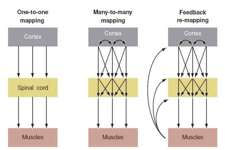
Mapping from motor cortex to muscles
- No one to one mapping
- Mnay-to-many mapping, and its modulated by feedback
Premotor and SMA
- Premotor stim can induce more complex movements than M1
- Premotor lesions, movements can be executed but obstacles can no longer be avoided
- Mental rehearsal activates premotor areas but not M1
M1 more involved in actual motor control
Premotor areas involved in motor planning
PM vs SMA
- Premotor more involved in sensory guided info
- Complex new movements
- SMA involved in pre-learned movement
- Muscle memory
Pathways
Dorsal/ventral split in motor cortex too
- Dorsal (where) -> In charge of reaching, culminates in PMd
- Ventral (what) -> In charge of grasping, culminates in PMv
Mirror Neurons
Activity in PMv that activates when monkey plansan action, but also when they see an action
Optimal Motor Control
Desired movement -> f_inv (f_inv(x) ) -> Muscle Activation -> Actual body ( f() ) -> Resulting movement
- Take a desired outcome x:
-> x is learned / trained. - Somatosensory function
Moving on from that:
- Harris & Wolpert proposes that optimal control theory relies on a few simple assumptions:
- Standard deviation of noise increases linearly with the absolute value of the control signal
- What matters is the variability at the end of the movement
- Additionally:
- Like we assumed, speed means more error
- But longer time means error can accumulate over time
Optimizing for speed and accuracy, but higher speed means higher variance
Week 8 - Cerebellum
// Review slides
Cerebellum
- Anatomy and functional division
- Anatomical structure of cerebellar cortex
- Cerebellar plasticity
- Overall roles of cerebellum
- Motor control: feedforward vs feedback controller
- Sensorimotor learning
- Timing
- Examples: eyeblink conditioning, saccade adaptation
Rough facts
-
10% of volume, more than half of neurons
-
40 times more axons project TO it than FROM it
-
Each purkinje cell (output neurons) receives input from approx 1 million granule
-
Arranged in highly regular manner as repeating units (basic circuit modules)
-
Can be divided into different regions, which perform similar computations based on different
-
Involved in the adjustment of movements while in progress and during repitition
-
Project to:
- Premotor and motor systems of cortex and brain stem
-
Cerebellar systems are ipsilateral to the ontrolled muscles (opposite to cortex)
-
Damage
- Disrupts spatial accuracy and temporal coordination of movements
- Imparis motor learning and certain cognitive function
-
Can be divided into the cerebellar cortex and 3 deep nuclei:
- Fastigial, interposed, and dentate nucleus
- Interposed split betweeen: globuse and emboliform nucleus
- Fastigial, interposed, and dentate nucleus
-
Output mainly originates from deep nuclei EXCEPT for flocculonodular lobe (projects directly to vestibular nuclei)
Anatomy
Functional division
- Flocculonodular lobe:
- vestibular functions
- vermis
- Receives multi-sensory inputs (sensory input from spinal cord)
- Involved in:
- Posture, locomotion, gaze control (via fastigial nucleus)
- Intermediate part of hemisphere:
- Somatosensory input from spinal cord
- Projects to spinal motor systems via the interposed nucleus
- Lateral part of the hemisphere
- Only cortical input
- Projects to motor, premotor, and PFC via the dentate nucleus
Anatomical structure of cerebellar cortex
- 3 layers
- 5 types of neurons (first four (all aside from granule) are inhibitory)
- Stellate (-)
- Basket (-)
- Purkinje (-)
- Golgi (-)
- Granule (+)
- Outer layer:
- Badies of stellate & basket cells
- Axons of granule cells (“parallel fibers”)
- Dendrites of Purkinje cells (perpendicular to parallel fibers)
- Purkinje cell layer
- Bodies of Purkinje cells
- Project to cerebellar/vestibular nuclei, output of cerebellar cortex
- Bodies of Purkinje cells
- Inner layer: granular layer
- Bodies of granule & golgi cells
- Mossy fibers (major input to cerebellum) terminate in this layer
Fibers:
- Mossy Fibers (input)
- Sensory?
- Major input to cerebellum, terminate on granular layer with granule and golgi cells
- Originate in: precerebellar nucleus cell (spinocerebellar pathways, brainstem reticular nuclei, pontine nuclei, etc.)
- Climbing Fibers (input)
- Error?
- Originate from inferior olive
- Parallel Fibers
- The axons of granule cells
- On the molecular layer, projecting between basket cells, stellate cells, and dendrites of Purkinje cells
Mossy Fibers -> Granule Cells -> Purkinje cells (PC)
- Purkinje receives input from only one climbing fiber
- Climbing fiber usually contacts up to 10 PC
- Synaptic effects of climbing fibers on PC are unusually powerful, producing complex spikes
- Activity in climbing fibers induces selective LTD in the synaptic strength of parallel fibers
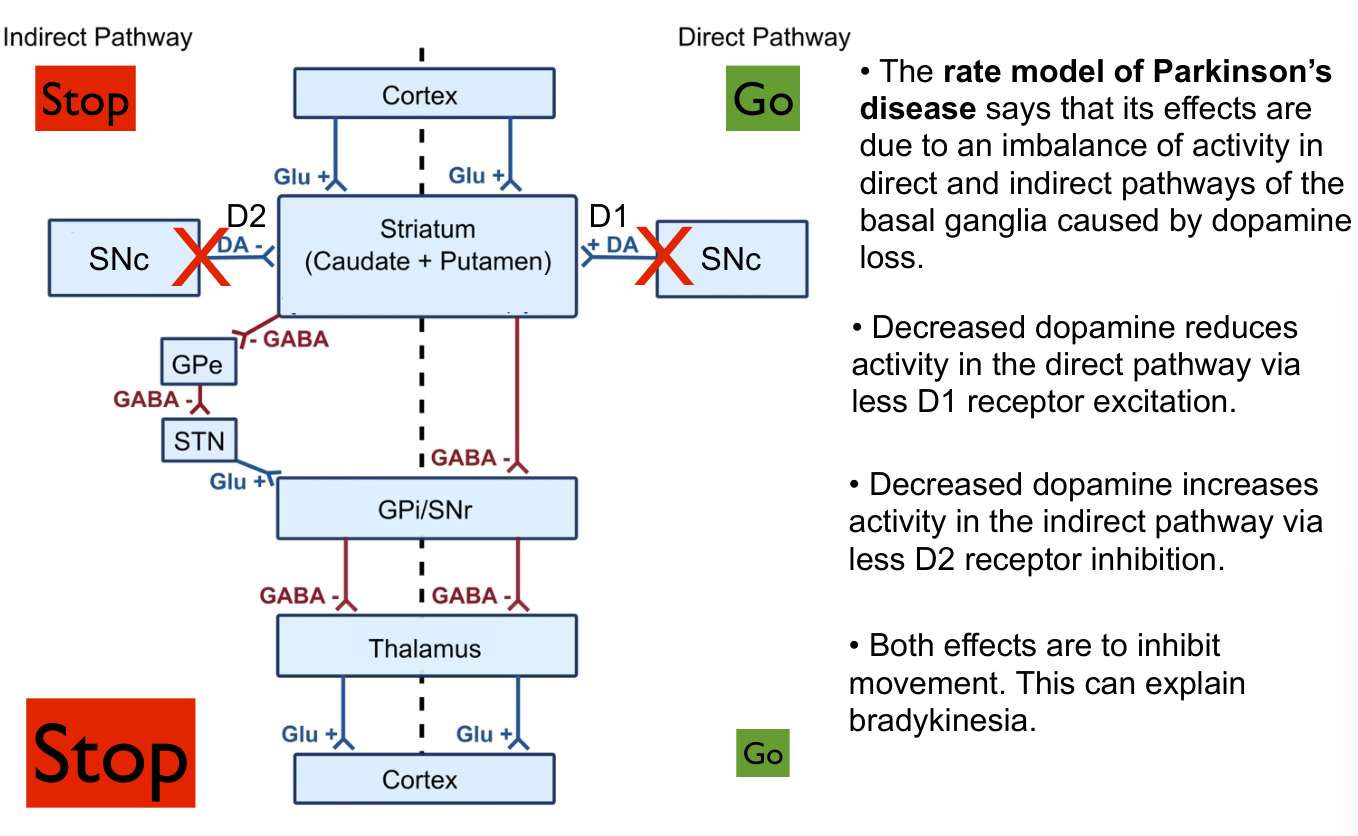
Week 9 - Basal Ganglia
// Review slides
Basal Ganglia
- Anatomy: 4 nuclei
- Mechanisms of actions (direct and indirect pathways)
- Huntington disease
- Parkinson disease and treatments
- Unifying theory: an action (or thought) selection network that is modified by reward-driven learning
- Cost-benefit dependent action selection model
Week 10 - Sensorimotor Integrations
No Summary but:
Know important cortical association areas
- Frontal Association Cortex
- Dorsal / Ventral (where/what) streams
- Dorsal:
- Parietal Association Cortex
- Somatosensory cortex
- Ventral
- Auditory Cortex
- Temporal Association cortex
- Neglect
- WM
- Decision Integrators
- Motion (MT)
- Relative stimulation frequence (somatosensory)
- Sequential probability test